Cardiovascular Imaging A handbook for clinical practice - Part 5 potx

Cardiovascular Imaging A handbook for clinical practice - Part 5 potx
... seg-
120 Chapter 10
Table 10.1 Imaging techniques: advantages and limitations.
Technique Advantage Limitation
Conventional angiography Gold standard Invasive
Risk
Cost
MRA Non-invasive Availability
Cost
CTA ... availability.
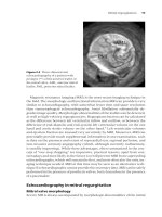
Magnetic resonance angiography
Gadolinium-enhanced magnetic resonance angiography (MRA) has proven
valuable for the non-invasive assessment of peripheral vascular di...

Cardiovascular Imaging A handbook for clinical practice - Part 1 pptx
... index.
ISBN-13: 97 8-1 -4 05 1-3 13 1 -5 (alk. paper)
ISBN-10: 1-4 05 1-3 13 1-4 (alk. paper)
1. Heart
—
Imaging
—
Handbooks, manuals, etc. 2. Cardiovascular system
—
Imaging
—
Handbooks, manuals, etc.
[DNLM: ... published 20 05
Library of Congress Cataloging-in-Publication Data
Cardiovascular imaging : a handbook for clinical practice / edited by Jeroen J....
Cardiovascular Imaging A handbook for clinical practice - Part 2 pps
... 3
CSA
LVOT
VTI
LVOT
VTI
As-Jet
Stenotic
valve
AVA
Figure 3.7 Schematic diagram of the continuity equation to calculate the aortic valve
area. AVA, aortic valve area; CSA, cross-sectional area; LVOT, left ventricular ... LVOT diameter measured from a parasternal long axis view for calculation of
a circular cross-sectional area.
2 LVOT velocity measured with pulsed wave Doppler from an ap...

Cardiovascular Imaging A handbook for clinical practice - Part 3 pptx
... the
mitral valve.
BCI7 6/18/ 05 11:17 AM Page 78
52 Chapter 5
Figure 5. 1 A 69-year-old man presented with acute back pain and diaphoresis.
Transesophageal echocardiography (TEE) showed acute type A aortic ... as-
cending aorta.
Echocardiography
Echocardiography (TTE and TEE) is a non-invasive and readily available
modality for evaluation of aortic dissection. TTE can evaluate th...

Cardiovascular Imaging A handbook for clinical practice - Part 4 docx
... tests that are available to clinicians are based
on the assessment of regional and global function, regional myocardial perfu-
sion, myocardial metabolism, or coronary anatomy. The cardiovascular ... pharmacologic
myocardial perfusion imaging that are associated with high-risk CAD and an
increased probability of future cardiac events.
A large amount of data published in the literature...

Cardiovascular Imaging A handbook for clinical practice - Part 6 doc
... coro-
nary artery bypass graft patency, and anomalous coronary arteries.
5
Acute dyspnea 157
Figure 13.2 CMR (left) and X-ray (right) coronary angiography in a patient with high-
grade proximal and ... diagnoses
Acute dyspnea 159
Figure 13.4 Apical three-chamber transthoracic echocardiogram in an 83-year-old
man with acute pulmonary edema 2 days after acute anterior myocardial infarctio...

Cardiovascular Imaging A handbook for clinical practice - Part 7 pot
... mechanical dyssynchrony and improves hemodynamics.
Conventional echocardiographic parameters
Table 15. 1 provides an overview on the available conventional parameters that
are valuable for the assessment ... integrity from parasternal and apical
long-axis as well as parasternal short-axis projections and these represent the
optimal views for the visualization and assessment of SAM.
There...

Cardiovascular Imaging A handbook for clinical practice - Part 8 pdf
... provides
improved imaging of smaller masses, particularly in the atria, atrial appendages,
or associated with valvular structures. Contrast-enhanced echocardiography
improves visualization of intracardiac masses ... symptoms.
Lipomas are typically located in the RA or atrial septum. They arise from the en-
docardial surface and have a broad base of attachment. Lipomas have the same
signal...

Cardiovascular Imaging A handbook for clinical practice - Part 9 pptx
... by 201 Tl-myocardial scintigraphy or abnormal accumulation by 67 Ga-
citrate or 99 m TC-myocardial scintigraphy
4. Abnormal intra-cardiac pressure, low cardiac output, or abnormal wall motion ... 6/18/ 05 11:09 AM Page 262
New imaging modalities
Magnetic resonance imaging
Magnetic resonance perfusion imaging (MRPI) using gadolinium-based con-
trast agents has recently been validated as...

Cardiovascular Imaging A handbook for clinical practice - Part 10 pps
... deformations may also be present. Catheter-driven angiography
has clear advantages in imaging quality, but radiation burden and invasiveness
are important drawbacks. Therefore, magnetic resonance imaging ... non-cardiac
risk stratification after 129– 35
in clinical decision-making 131–3
rest imaging 129–30
stress imaging 130–1
myocardial ischemia
acute, non-invasive imaging 156 –8...
Từ khóa:
- academic writing a handbook for international students
- a handbook for medical teachers newble
- academic writing a handbook for international students answers
- academic writing a handbook for international students by stephen bailey
- academic writing a handbook for international students 3rd edition answers
- academic writing a handbook for international students 3rd edition
- Báo cáo thực tập tại nhà thuốc tại Thành phố Hồ Chí Minh năm 2018
- chuyên đề điện xoay chiều theo dạng
- Nghiên cứu tổ chức pha chế, đánh giá chất lượng thuốc tiêm truyền trong điều kiện dã ngoại
- Một số giải pháp nâng cao chất lượng streaming thích ứng video trên nền giao thức HTTP
- Biện pháp quản lý hoạt động dạy hát xoan trong trường trung học cơ sở huyện lâm thao, phú thọ
- Giáo án Sinh học 11 bài 13: Thực hành phát hiện diệp lục và carôtenôit
- Giáo án Sinh học 11 bài 13: Thực hành phát hiện diệp lục và carôtenôit
- ĐỒ ÁN NGHIÊN CỨU CÔNG NGHỆ KẾT NỐI VÔ TUYẾN CỰ LY XA, CÔNG SUẤT THẤP LPWAN
- ĐỒ ÁN NGHIÊN CỨU CÔNG NGHỆ KẾT NỐI VÔ TUYẾN CỰ LY XA, CÔNG SUẤT THẤP LPWAN
- Quản lý hoạt động học tập của học sinh theo hướng phát triển kỹ năng học tập hợp tác tại các trường phổ thông dân tộc bán trú huyện ba chẽ, tỉnh quảng ninh
- Phối hợp giữa phòng văn hóa và thông tin với phòng giáo dục và đào tạo trong việc tuyên truyền, giáo dục, vận động xây dựng nông thôn mới huyện thanh thủy, tỉnh phú thọ
- Phát triển mạng lưới kinh doanh nước sạch tại công ty TNHH một thành viên kinh doanh nước sạch quảng ninh
- Nghiên cứu về mô hình thống kê học sâu và ứng dụng trong nhận dạng chữ viết tay hạn chế
- Nghiên cứu tổng hợp các oxit hỗn hợp kích thƣớc nanomet ce 0 75 zr0 25o2 , ce 0 5 zr0 5o2 và khảo sát hoạt tính quang xúc tác của chúng
- Kiểm sát việc giải quyết tố giác, tin báo về tội phạm và kiến nghị khởi tố theo pháp luật tố tụng hình sự Việt Nam từ thực tiễn tỉnh Bình Định (Luận văn thạc sĩ)
- BT Tieng anh 6 UNIT 2
- Tranh tụng tại phiên tòa hình sự sơ thẩm theo pháp luật tố tụng hình sự Việt Nam từ thực tiễn xét xử của các Tòa án quân sự Quân khu (Luận văn thạc sĩ)
- chuong 1 tong quan quan tri rui ro
- Giáo án Sinh học 11 bài 14: Thực hành phát hiện hô hấp ở thực vật
- Giáo án Sinh học 11 bài 14: Thực hành phát hiện hô hấp ở thực vật