báo cáo khoa học: " Surgical treatment of giant mesenteric fibromatosis presenting as a gastrointestinal stromal tumor: a case report" doc
báo cáo khoa học: " Surgical treatment of giant mesenteric fibromatosis presenting as a gastrointestinal stromal tumor: a case report" doc
... 11:2373-2379.
doi:10.1186/1752-1947-4-314
Cite this article as: Stoidis et al.: Surgical treatment of giant mesenteric
fibromatosis presenting as a gastrointestinal stromal tumor: a case
report. Journal of Medical Case Reports 2010 ... Case presentation
A 64-year-old Caucasian man was admitted to our hos-
pital with a ten- year-history of a mild diff...

báo cáo khoa học: "Surgical treatment of intracystic carcinoma of the breast" doc
... Matsuda: matsuyo@asahikawa-med.ac.jp
Kazuhiro Sato: kiki@asahikawa-med.ac.jp
Naoyuki Miyokawa: mao@asahikawa-med.ac.jp
Tadahiro Sasajima: sasajima@asahikawa-med.ac.jp
Contact information of ... Asahikawa Medical University,
Asahikawa,
Hokkaido, Japan.
E-mail addresses:
Masasahiro Kitada: k1111@asahikawa-med.ac.jp
Satoshi Hayashi: shayashi@asahikawa-med.ac.jp
Yoshinari Mats...
Báo cáo khoa học: "Surgical management of giant Brunner''''s gland hamartoma: case report and literature review" ppsx
... purposes)
World Journal of Surgical Oncology
Open Access
Case report
Surgical management of giant Brunner's gland hamartoma: case
report and literature review
Zoe A Stewart
1
, Ralph H Hruban
2
, Elliot ... with a Giant
Brunner's Gland Hamartoma.
Histopathological Appearance of a Giant Brunner's Gland Hamartoma stained with Hematoxylin and Eosin and...

Báo cáo khoa học: "Surgical treatment of a rare primary renal carcinoid tumor with liver metastasis" docx
... tumor, with emphasis on the treatment of liver
metastases.
Case presentation
We evaluated a 45 year-old patient who presented initially
with abdominal pain. Abdominal and pelvic CT scan
showed lesions ... breasts, ovaries, testes, prostate and other
locations. We report a case of a carcinoid of renal origin with synchronous single liver metastases
on radiological studies.
C...
Báo cáo y học: "Surgical treatment of giant plexiform neurofibroma associated with pectus excavatum" pptx
... excava-
tum(PE)[4,5],whichisthemostcommonchestwall
malformation and one of t he most frequent major con-
genital anomalies. Here, we present a case of giant chest
wall PNF associated with PE.
2. Case Report
A 12-years-old boy ... Whether this association of pectus excava-
tum is a manifestation of the giant tumor or an inciden-
tal finding is unclear based on our case....

báo cáo khoa học: " Surgical treatment of gingival overgrowth with 10 years of follow-up" potx
... was carried out with manual and
ultrasonic instruments to complete the baseline therapy.
This prot ocol was able to eliminate the local aggrava-
tion factors and thus guarantee a good surgical ... had an
aesthetically satisfactory gingival appearance and no sign
of recurrence.
All the grafts were well vascularized and aesthetically
satisfactory.
Unlike the classic approach,thesurgicalte...
báo cáo khoa học: " Preclinical evaluation of KIT/PDGFRA and mTOR inhibitors in gastrointestinal stromal tumors using small animal FDG PET" ppsx
... Isozaki K, Moriyama Y, Hashimoto K, Nishida T, Ishiguro S,
Kawano K, Hanada M, Kurata A, Takeda M, Muhammad Tunio G,
Matsuzawa Y, Kanakura Y, Shinomura Y, Kitamura Y: Gain of function
mutations of ... tumor mass was
evaluated using small animal PET tomography in one
animal per group (37 days after cell injection). The
base-line FDG uptake was p ositive in all animals evalu-
ated with a...
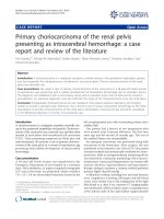
báo cáo khoa học: " Primary choriocarcinoma of the renal pelvis presenting as intracerebral hemorrhage: a case report and review of the literature" docx
... lesions at the area of the hematoma, a
metastatic lesion situated on her left temporal lobe,
whereas a magnetic angiography did not reveal any
vascular dysplasia.
On the 26
th
day of her ICU stay, ... syncytiotrophoblasts and trophoblasts in a
carcinomatous area (hematoxylin and eosin stain; magnification ×
200).
Kyriakou et al. Journal of Medical Case Reports 2011, 5:501
http:/...
Từ khóa:
- báo cáo khoa học mẫu
- báo cáo khoa học y học
- báo cáo khoa học sinh học
- báo cáo khoa học nông nghiệp
- báo cáo khoa học lâm nghiệp
- báo cáo khoa học thủy sản
- Báo cáo thực tập tại nhà thuốc tại Thành phố Hồ Chí Minh năm 2018
- Báo cáo quy trình mua hàng CT CP Công Nghệ NPV
- chuyên đề điện xoay chiều theo dạng
- Nghiên cứu tổ chức pha chế, đánh giá chất lượng thuốc tiêm truyền trong điều kiện dã ngoại
- đề thi thử THPTQG 2019 toán THPT chuyên thái bình lần 2 có lời giải
- Giáo án Sinh học 11 bài 13: Thực hành phát hiện diệp lục và carôtenôit
- Giáo án Sinh học 11 bài 13: Thực hành phát hiện diệp lục và carôtenôit
- Giáo án Sinh học 11 bài 13: Thực hành phát hiện diệp lục và carôtenôit
- ĐỒ ÁN NGHIÊN CỨU CÔNG NGHỆ KẾT NỐI VÔ TUYẾN CỰ LY XA, CÔNG SUẤT THẤP LPWAN
- NGHIÊN CỨU CÔNG NGHỆ KẾT NỐI VÔ TUYẾN CỰ LY XA, CÔNG SUẤT THẤP LPWAN SLIDE
- Phối hợp giữa phòng văn hóa và thông tin với phòng giáo dục và đào tạo trong việc tuyên truyền, giáo dục, vận động xây dựng nông thôn mới huyện thanh thủy, tỉnh phú thọ
- Phát triển mạng lưới kinh doanh nước sạch tại công ty TNHH một thành viên kinh doanh nước sạch quảng ninh
- Phát triển du lịch bền vững trên cơ sở bảo vệ môi trường tự nhiên vịnh hạ long
- Phát hiện xâm nhập dựa trên thuật toán k means
- Thiết kế và chế tạo mô hình biến tần (inverter) cho máy điều hòa không khí
- Sở hữu ruộng đất và kinh tế nông nghiệp châu ôn (lạng sơn) nửa đầu thế kỷ XIX
- Kiểm sát việc giải quyết tố giác, tin báo về tội phạm và kiến nghị khởi tố theo pháp luật tố tụng hình sự Việt Nam từ thực tiễn tỉnh Bình Định (Luận văn thạc sĩ)
- Quản lý nợ xấu tại Agribank chi nhánh huyện Phù Yên, tỉnh Sơn La (Luận văn thạc sĩ)
- Tranh tụng tại phiên tòa hình sự sơ thẩm theo pháp luật tố tụng hình sự Việt Nam từ thực tiễn xét xử của các Tòa án quân sự Quân khu (Luận văn thạc sĩ)
- Giáo án Sinh học 11 bài 14: Thực hành phát hiện hô hấp ở thực vật