Báo cáo khoa học: X-ray structure of glucose/galactose receptor from Salmonella typhimurium in complex with the physiological ligand, (2R)-glyceryl-b-D-galactopyranoside pdf
Báo cáo khoa học: X-ray structure of glucose/galactose receptor from Salmonella typhimurium in complex with the physiological ligand, (2R)-glyceryl-b-D-galactopyranoside pdf
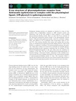
... protein. This kind of protein is exemplified by the
Thermus thermophilus protein, PDB entry 2B3B [28].
The mode of binding the monosaccharide is completely
different in terms of orientation of the ... calcium site of domain 2 was described
earlier, and tight binding of the ion was shown to con-
tribute to the integrity of the protein structure [17,22].
The sod...
Báo cáo khoa học: Crystal structure of a designed tetratricopeptide repeat module in complex with its peptide ligand pot
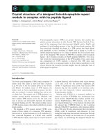
... conformation. At the N-terminus of the
peptide, the alignment diverges more, with the major
difference being that the N-terminal Met is signifi-
cantly further away from the binding cleft in the
CTPR390–MEEVD ... complexes are in
the location of the peptide chains relative to the pro-
tein (Fig. 5A). The Hsp90 peptide is located in the
CTPR390 concave clef...
Báo cáo khoa học: Crystal structure of RNase A tandem enzymes and their interaction with the cytosolic ribonuclease inhibitor potx
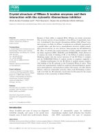
... calculations
for the two interacting molecular surfaces, performed
with the program sc from the ccp4 suite [27]. The
obtained sc value of 0.400 indicates weak binding of
the two entities of the RATE. Therefore, ... of RNase molecules
on the cell surface), thereby favoring their endocytosis
[19]. The stoichiometry of RI binding to RATEs and
the role of the R...

Tài liệu Báo cáo khoa học: Crystal structure of Trypanosoma cruzi glyceraldehyde-3-phosphate dehydrogenase complexed with an analogue of 1,3-bisphospho-D-glyceric acid Selective inhibition by structure-based design docx
... elsewhere on the
pathway of the multistep catalysis, where the OH inter-
actions with residues of the active site are not required.
Using information on the 3D structure of the enzyme–
inhibitor complex, ... structures [24]. Parallel studies of these effects
allowed identification of the specific interactions between
the inhibitors and the proteins. In the a...
Báo cáo khoa học: Solution structure of Cu6 metallothionein from the fungus Neurospora crassa pptx
... data. The accu-
racy of the structure calculated without these constraints is,
however, supported by the similarities of the 800 MHz
structures of the a-domain of mouse MT1 compared to the
one ... to the average minimized structure,
for each set of reintroduced long-range NOEs. Determining
the deviation of the generated structure with a reduced
number of...

Tài liệu Báo cáo khoa học: Coordination chemistry of iron(III)±porphyrin±antibody complexes In¯uence on the peroxidase activity of the axial coordination of an imidazole on the iron atom ppt
... The bind-
ing of the ®rst CN
±
ligand to the iron was thus more easy in
the hydrophobic binding pocket of the antibody than the
binding of the second one, most probably because of the
steric hindrance ... substituents
of the meso-phenyl rings being recognized by th e side chains
of amino acids of the antibody [36]; (b) in the case of 13G10,
one carbox...
Báo cáo khoa học: "Minimalist Parsing of Subjects Displaced from Embedded Clauses in Free Word Order Languages" ppt
... those in the “input queue,” and
if not, shifting a word from the input queue onto the
processing buffer. The distinction is marked, in our
notation, by a |: the words and trees before | are in
the ... if
there is another displaced item in the tree containing
the original CH that is compatible with the UNCH
feature but displaced from some other phrase. This
requ...

Tài liệu Báo cáo khoa học: Crystal structure of an ascomycete fungal laccase from Thielavia arenaria – common structural features of asco-laccases ppt
... gives the solvent
access to the trinuclear centre. However, the channel is
blocked by the C-terminal end of the amino acid chain
in MaL ⁄rMaL [25,33]. Similarly, in the structure of
TaLcc1, the ... DSGL as the last four amino acids
penetrating into the channel.
The C-terminal processing has been reported for
asco-laccases of different origins [37–39]; furthermo...

Tài liệu Báo cáo khoa học: Crystal structure of importin-a bound to a peptide bearing the nuclear localisation signal from chloride intracellular channel protein 4 ppt
... determined the X-ray crystal structure of a truncated form
of importin-a lacking the importin-b binding domain, bound to a CLIC4
NLS peptide. The NLS peptide binds to the major binding site in an
extended ... cargo binding sites, referred to as
the major and minor binding sites [1]. These sites are
located in the concave face of the protein near regions
of invari...

Tài liệu Báo cáo khoa học: Crystal structure of the cambialistic superoxide dismutase from Aeropyrum pernix K1 – insights into the enzyme mechanism and stability pdf
... metal-binding site
at the interface of the two domains, which consists
of four side chains: two (His31 and His79) from the
N-terminal domain and two (Asp165 and His169) from
the C-terminal domain ... focus
on the coordination of the metal cofactor in the active
site as well as the changes it experiences in response to
different metal cofactors. Finally, by compar...
Từ khóa:
- báo cáo khoa học mẫu
- báo cáo khoa học y học
- báo cáo khoa học sinh học
- báo cáo khoa học nông nghiệp
- báo cáo khoa học lâm nghiệp
- báo cáo khoa học thủy sản
- Báo cáo quy trình mua hàng CT CP Công Nghệ NPV
- chuyên đề điện xoay chiều theo dạng
- Nghiên cứu tổ chức pha chế, đánh giá chất lượng thuốc tiêm truyền trong điều kiện dã ngoại
- Nghiên cứu tổ hợp chất chỉ điểm sinh học vWF, VCAM 1, MCP 1, d dimer trong chẩn đoán và tiên lượng nhồi máu não cấp
- Nghiên cứu tổ chức chạy tàu hàng cố định theo thời gian trên đường sắt việt nam
- Biện pháp quản lý hoạt động dạy hát xoan trong trường trung học cơ sở huyện lâm thao, phú thọ
- Giáo án Sinh học 11 bài 13: Thực hành phát hiện diệp lục và carôtenôit
- Giáo án Sinh học 11 bài 13: Thực hành phát hiện diệp lục và carôtenôit
- Quản lý hoạt động học tập của học sinh theo hướng phát triển kỹ năng học tập hợp tác tại các trường phổ thông dân tộc bán trú huyện ba chẽ, tỉnh quảng ninh
- Phát hiện xâm nhập dựa trên thuật toán k means
- Nghiên cứu, xây dựng phần mềm smartscan và ứng dụng trong bảo vệ mạng máy tính chuyên dùng
- Nghiên cứu tổng hợp các oxit hỗn hợp kích thƣớc nanomet ce 0 75 zr0 25o2 , ce 0 5 zr0 5o2 và khảo sát hoạt tính quang xúc tác của chúng
- Nghiên cứu khả năng đo năng lượng điện bằng hệ thu thập dữ liệu 16 kênh DEWE 5000
- Tìm hiểu công cụ đánh giá hệ thống đảm bảo an toàn hệ thống thông tin
- Quản lý nợ xấu tại Agribank chi nhánh huyện Phù Yên, tỉnh Sơn La (Luận văn thạc sĩ)
- Tăng trưởng tín dụng hộ sản xuất nông nghiệp tại Ngân hàng Nông nghiệp và Phát triển nông thôn Việt Nam chi nhánh tỉnh Bắc Giang (Luận văn thạc sĩ)
- Giáo án Sinh học 11 bài 15: Tiêu hóa ở động vật
- chuong 1 tong quan quan tri rui ro
- Giáo án Sinh học 11 bài 14: Thực hành phát hiện hô hấp ở thực vật
- Giáo án Sinh học 11 bài 14: Thực hành phát hiện hô hấp ở thực vật