Báo cáo khoa học: NMR structure of the thromboxane A2 receptor ligand recognition pocket pot

Báo cáo khoa học: NMR structure of the thromboxane A2 receptor ligand recognition pocket pot
... further definition of the ligand recognition
pocket on the extracellular side of the receptors. This
has become a major obstacle to the further understanding
of the molecular mechanism of the ... conformation
change of the receptor and triggering the coupling of the
receptor with the G protein in the intracellular domain s.
The first step of bindi...

Tài liệu Báo cáo khoa học: NMR structure of AII in solution compared with the X-ray structure of AII bound to the mAb Fab131 pptx
... (B) structure alignment of
the fragment 4–7 of the 13 ensemble structures of AII to the fragment 3–6
of the X-ray structure of AII.
Fig. 6. Structure of a representative folded conformer of AII ... Lennard-
Jones potentials are used. From the family of the 48
structures, 13 structures were selected having the best
allowed regions in the Ramachandran plot...

Tài liệu Báo cáo khoa học: Crystal structure of the cambialistic superoxide dismutase from Aeropyrum pernix K1 – insights into the enzyme mechanism and stability pdf
... [26]. These findings
lead to the hypothesis that Fe-bound ApeSOD mimics
the product-inhibited form and the shift of Tyr39 sup-
presses the release of the peroxide product. This may
be one of the ... the ter-
tiary structure of ApeSOD has not been elucidated.
In the present study, for the first time, we describe the
crystal structure of ApeSOD. In particular, we...

Tài liệu Báo cáo khoa học: Crystal structure of the catalytic domain of DESC1, a new member of the type II transmembrane serine proteinase family pptx
... Lys224–
Tyr228 (the back of the pocket) and the disulfide bridge
Cys191–Cys220 (the front of the pocket) (Fig. 4A). The
backbones of these segments form a deep hydrophobic
pocket with the negatively ... according to the residue in the mid-
point of the respective loop, as shown in Fig. 2. To
the east of the active site the 37- and 60-loops border
the S2¢...

Tài liệu Báo cáo khoa học: Crystal structure of the BcZBP, a zinc-binding protein from Bacillus cereus doc
... corresponding to the
N-terminus of the a2- helix and to the preceding loop
results in a rmsd of 1.0 A
˚
for the Ca atoms. Thus,
the movement of the a2 helix accounts for 23% of the
rmsd value (i.e. for ... determines the shape of the
active site entry. (e) The structure of the active site is
essentially identical with the active sites of the MshB
a...
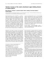
Tài liệu Báo cáo khoa học: Solution structure of the matrix attachment region-binding domain of chicken MeCP2 ppt
... one-turn helix on the other face. It is thought
that the two inner strands of the b-sheet lie within the major
groove of the DNA and that a hydrophobic pocket formed
by the side chains of Y123 and ... [18]. The structure of the helical
coil a
2
/a
3
allows us to interpret the consequences of the
six mutations. As P153(152) and G162(161) are buried in
the pr...
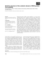
Báo cáo khoa học: Solution structure of the catalytic domain of RICH protein from goldfish pot
... regrowth
upon the optic nerve crush, and also expressed in the
germinal neuroepithelium of retina, which generates
new neurons throughout the lifespan of the fish [11].
The cloning of the RICH proteins ... structures. The lowest-energy structure from the
RICH NMR ensemble is used for the overlay. (D) The surface of the RICH catalytic domain shows several negat...

Báo cáo khoa học: Solution structure of the active-centre mutant I14A of the histidinecontaining phosphocarrier protein from Staphylococcus carnosus ppt
... towards the
interior of the protein. The space that in the wild-type
molecule is occupied by the large hydrophobic side chain of
Fig. 3. Structure ensemble of HPr(I14A). The average structure of the
10 ... was
computed as the ratio of the s tandard deviations of the
chemical shifts of the amide nitrogen and p roton nuclei.
Results
Determination of th...
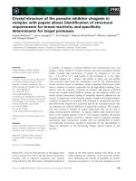
Báo cáo khoa học: Crystal structure of the parasite inhibitor chagasin in complex with papain allows identification of structural requirements for broad reactivity and specificity determinants for target proteases pptx
... part, and (b) the variability of the angle of
approach of the inhibitor relative to the catalytic
cleft of the enzyme. The latter factor may reflect not
so much the geometry of the catalytic site ... PW),
despite the lack of overall sequence similarity. The
role of the proline residue appears to be to maintain
the specific shape of the loop. The aroma...
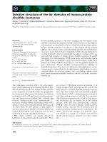
Báo cáo khoa học: Solution structure of the bb¢ domains of human protein disulfide isomerase docx
... relative
orientations of the a and b domains. In one struc-
ture, the catalytic cysteines face each other; in the
other, the catalytic residues of the a domain face
away from the a¢ domain. The crystal structures ... mm of
monomers at 30 °C. Most of the
1
H-
15
N HSQC signals
of the dimeric form of PDI-bb¢ coincide with the signals
of the monomeric form or...
Từ khóa:
- Báo cáo thực tập tại nhà thuốc tại Thành phố Hồ Chí Minh năm 2018
- Báo cáo quy trình mua hàng CT CP Công Nghệ NPV
- Nghiên cứu sự hình thành lớp bảo vệ và khả năng chống ăn mòn của thép bền thời tiết trong điều kiện khí hậu nhiệt đới việt nam
- Nghiên cứu vật liệu biến hóa (metamaterials) hấp thụ sóng điện tử ở vùng tần số THz
- đề thi thử THPTQG 2019 toán THPT chuyên thái bình lần 2 có lời giải
- Giáo án Sinh học 11 bài 13: Thực hành phát hiện diệp lục và carôtenôit
- Giáo án Sinh học 11 bài 13: Thực hành phát hiện diệp lục và carôtenôit
- ĐỒ ÁN NGHIÊN CỨU CÔNG NGHỆ KẾT NỐI VÔ TUYẾN CỰ LY XA, CÔNG SUẤT THẤP LPWAN
- Phát triển mạng lưới kinh doanh nước sạch tại công ty TNHH một thành viên kinh doanh nước sạch quảng ninh
- Phát triển du lịch bền vững trên cơ sở bảo vệ môi trường tự nhiên vịnh hạ long
- Phát hiện xâm nhập dựa trên thuật toán k means
- Nghiên cứu về mô hình thống kê học sâu và ứng dụng trong nhận dạng chữ viết tay hạn chế
- Nghiên cứu khả năng đo năng lượng điện bằng hệ thu thập dữ liệu 16 kênh DEWE 5000
- Thơ nôm tứ tuyệt trào phúng hồ xuân hương
- Chuong 2 nhận dạng rui ro
- Tổ chức và hoạt động của Phòng Tư pháp từ thực tiễn tỉnh Phú Thọ (Luận văn thạc sĩ)
- Kiểm sát việc giải quyết tố giác, tin báo về tội phạm và kiến nghị khởi tố theo pháp luật tố tụng hình sự Việt Nam từ thực tiễn tỉnh Bình Định (Luận văn thạc sĩ)
- Tăng trưởng tín dụng hộ sản xuất nông nghiệp tại Ngân hàng Nông nghiệp và Phát triển nông thôn Việt Nam chi nhánh tỉnh Bắc Giang (Luận văn thạc sĩ)
- Giáo án Sinh học 11 bài 15: Tiêu hóa ở động vật
- Giáo án Sinh học 11 bài 14: Thực hành phát hiện hô hấp ở thực vật